


Sacroiliac Joint Arthrodesis
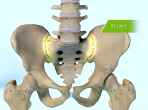
Background – The sacroiliac (SI) joint is an often-overlooked source of low back pain. The sacroiliac joint is where the low back (sacrum) meets the hips (iliac crest). Studies estimate that the SI joint is responsible for up to 30% of chronic low back pain and up to 40% of chronic low back pain following lumbar fusion. Pain from the SI joint can refer to the hips, to the groin, or down the leg like sciatica. The SI joint acts as a capstone between the low back and the pelvis. The joint is reinforced with strong ligaments so there is typically very little motion at the SI joint. Risk factors for sacroiliac joint dysfunction include pelvic changes after pregnancy and childbirth and change in biomechanics and force load after lumbar spine fusion.
Symptoms of Sacroiliac joint dysfunction – Like any other joint in the body, the SI joint can be injured and/or undergo degeneration. When this happens, people can feel pain in their buttock and sometimes in the lower back, hips and legs. This is especially true while lifting, running, walking or even lying on the involved side.
A dysfunctional SI joint may inflame the joint and surrounding nerves causing pain in the lower back, hip, groin, or pelvis.
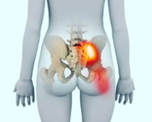
It’s common for pain from the SI joint to feel like disc or lower back pain, or sometimes hip or groin pain. For this reason, SI joint disorders should always be considered in lower back, hip, and pelvic pain diagnosis.
Do you experience one or more of the symptoms listed below?
- Lower back pain
- Sensation of low extremity: pain, numbness, tingling, weakness
- Pelvis/buttock pain
- Hip/groin pain
- Feeling of leg instability (buckling, giving way)
- Disturbed sleep patterns due to pain
- Disturbed sitting patterns (unable to sit for long periods, sitting on one side)
- Pain going from sitting to standing

Patients who suffer from SI joint dysfunction can have severe pain when performing transitional movements like standing from a chair.

Patients who have SI joint pain usually find it difficult to sit for long periods of time, and usually try to alleviate the discomfort by sitting on the least effected side.
Making a Diagnosis – A variety of tests performed during physical examination may help reveal the SI joint as the cause of your symptoms. Sometimes, X-rays, CT-scan or MRI may be helpful in the diagnosis of SI joint-related problems because they can rule out other common sources of pain—such as your lumbar spine or hip joints. It is also important to remember that other conditions (like a disc problem) can co-exist with SI joint disorders.
The most relied upon method to accurately determine whether the SI joint is the cause of your lower back pain symptoms is to inject the SI joint with a local anesthetic. This diagnostic injection will be performed under X-ray guidance to verify accurate placement of the needle in the SI joint. If your symptoms decrease by at least 75%, it can be concluded that the SI joint is either the source of or a major contributor to your lower back, hip, or pelvic pain. If the level of pain does not change after SI joint injection, it is less likely that the SI joint is the cause of your pain.
Treatment Options – Once the SI joint is confirmed as the cause of your symptoms, treatment can begin. Some patients respond well to physical therapy, use of oral medications, injection therapy, or radiofrequency ablation. These treatments are often performed repetitively, and frequently symptom improvement using these therapies is temporary. If non-surgical treatment options have been tried and do not provide long-term relief, it is time to consider minimally invasive sacroiliac joint arthrodesis.
Sacroiliac Joint Arthrodesis – If pain from the SI joint has not responded to conservative management or injection therapy and is significantly impacting function and quality of life, it is time to consider SI joint arthrodesis. Minimally-invasive SI joint arthrodesis involves implanting small screws across the SI joint to stabilize the joint and stop excessive motion. The procedure is fluoroscopically (XR) guided so there is only a small incision and a relatively short recovery period.
Sacroiliac joint arthrodesis stabilizes the SI joint. The procedure typically involves inserting three threaded titanium implants across the SI joint to maximize stability, reduce pain, and improve function. The procedure is done through a small one-inch incision and takes about an hour. The 3D-printed implant was designed for osseointegration, which is the structural and functional connection between implant and bone. This allows your painful joint to be stabilized through binding of bone all along the implant.

The 3D printed surface of the implant is similar to cancellous bone.
More than 100, peer-reviewed publications demonstrate the safety, durable effectiveness, and biomechanical and economic benefits of minimally invasive sacroiliac joint arthrodesis (www.si-bone.com/results). Multiple prospective clinical studies, including two randomized controlled trials, demonstrate that treatment of sacroiliac joint dysfunction with arthrodesis improved pain, patient function, and quality of life. As with any minimally invasive surgical procedures, there are potential risks associated with the procedure. It may not be appropriate for all patients and all patients may not benefit.
References
Polly DW, et al., and the INSITE Study Group. Two-Year Outcomes from a Randomized Controlled Trial of Minimally Invasive Sacroiliac Joint Fusion vs. Non-Surgical Management for Sacroiliac Joint Dysfunction. Int J Spine Surg. 2016;10:Article 28. DOI: 10.14444/3028
Dengler J, et al. Randomized Trial of Sacroiliac Joint Fusion vs. Conservative Management for Chronic Low Back Pain Attributed to the Sacroiliac Joint. J Bone Joint Surg Am. 2019;101(5):400-11. DOI: 10.2106/JBJS.18.00022.
Duhon B, Bitan F, Lockstadt H, Kovalsky D, Cher D, Hillen T, on behalf of the SIFI Study Group. Triangular Titanium Implants for Minimally Invasive Sacroiliac Joint Fusion: 2-Year Follow-Up from a Prospective Multicenter Trial. Int J Spine Surg. 2016;10:Article 13. DOI: 10.14444/3013
Dengler J, et al. on behalf of the INSITE, iMIA and SIFI study groups. Predictors of Outcome in Conservative and Minimally Invasive Surgical Management of Pain Originating from the Sacroiliac Joint – a Pooled Analysis. Spine. 2017;42(21):1664-73. [Epub 2017 Mar 27]. DOI: 10.1097/BRS.0000000000002169
Whang PG, et al. Long-Term Prospective Clinical and Radiographic Outcomes After Minimally Invasive Lateral Transiliac Sacroiliac Joint Fusion Using Triangular Titanium Implants. Med Devices (Auckl). 2019;12:411-422. DOI: 10.2147/MDER.S219862
Patel V, et al. Prospective Trial of Sacroiliac Joint Fusion Using 3D-Printed Triangular Titanium Implants: 24-Month Follow-Up. Med Devices (Auckl). 2021;14:211-216. DOI: 10.2147/MDER.S314828